Densitometry. X-ray densitometry What is densitometry
Densitometry is the main method for diagnosing early osteoporosis. This study is aimed at the central and peripheral parts of the skeleton, depending on the tasks assigned to the specialist.
Densitometers for the study of the axial skeleton - the spine and femurs (such densitometry is also called central) are stationary devices with an ultra-low dose x-ray tube that does not require the same security measures as x-ray machines. The radiation dose used in modern densitometric systems is 1000 times lower than with conventional X-rays.
Densitometry of the lumbar spine and femoral neck is taken as a basis. The study of these two zones is sufficient to obtain complete information about the state of the cortical and trabecular layers of the skeleton, as well as to assess the mineral density of bone tissue. Peripheral densitometry is performed as a screening study or when other parts of the skeleton are unavailable.
Also, peripheral examination is indicated in the presence of certain diseases that are sensitive to the effects of x-rays, for example, hyperparathyroidism. During such a study, a portable device is used, which gives a minimum of radiation and X-ray sensitive areas are removed to the maximum from the examination area.
When examining the lumbar spine, the vertebrae L1-L4 are taken in direct projection. If possible, all vertebrae that can be assessed should be examined. If it is impossible to diagnose all the vertebrae, densitometry and other zones should be added.
Densitometry of the femoral neck is performed to assess the condition of the cortical and spongy layer. For research, the proximal femur is taken. To clarify the results, the doctor can diagnose the Ward's area and the greater trochanter, which are not used in isolation in conventional densitometry.
Indications
As practice shows, its early diagnosis is extremely important for the successful treatment of osteoporosis. According to statistics, after the development of the first pathological fracture, the risk of developing repeated ones increases five times. Moreover, the fracture site will not depend on the location of the primary fracture. Accordingly, every specialist should know well who is at risk for osteoporosis. Doctors of any specialty can face this pathology: gynecologists, endocrinologists, gastroenterologists, neurologists, vertebrologists, rheumatologists, surgeons, traumatologists.
- Annual densitometry of the femoral neck and lumbar spine is recommended for every woman over 65 and every man over 70;
- If the patient is at risk, then densitometry is performed regardless of age.
Risk groups for osteoporosis:
- Patients taking steroids, corticosteroids, non-steroidal anti-inflammatory drugs;
- Patients receiving hormone therapy;
- Patients with insufficient body weight;
- Obese patients;
- Patients with endocrine pathology;
- Patients with rheumatic diseases;
- Patients with a history of low-traumatic fractures;
- Patients with relatives with osteoporosis.
Densitometry is also performed in patients with a suspected compression fracture. Plain x-ray can not always confirm the presence of such fractures, unlike densitometry.
And, of course, this examination is carried out to monitor the effectiveness of the treatment and for the screening diagnosis of osteoporosis. In addition to stating the fact of the development of osteoporosis, according to the results of the study, the doctor makes a forecast about the course of the disease, about the likelihood of developing pathological fractures.
Price
The price of densitometry in private clinics in Moscow can vary significantly: from a thousand to five or six thousand. The cost may depend on the study area: in some cases, densitometry of the entire skeleton is performed, on the type of study: ultrasound, X-ray, computer.
Med-7 has a modern X-ray densitometer. The cost of the examination is 2,200 rubles for a comprehensive study and 1,400 rubles for one zone, in terms of time, the diagnosis of one zone takes about 15 minutes.
The service includes:
- Study
- Snapshot disc
- Film sold separately
- Decoding by doctor radiologist
(impaired mineralization with a normal volume of bone tissue) and osteoporosis (decrease in density due to a decrease in bone mass). Osteopenia is quite often observed during pregnancy and lactation, so ultrasound densitometry is indispensable for examining women who are carrying and nursing a child. Despite the fact that the dose of ionizing radiation from X-ray densitometry is only a tenth of the radiation received from chest X-ray, even such a radiation exposure is undesirable for expectant and young mothers.
Osteoporosis is more of an age-related disease. In this regard, ultrasonic densitometry is indicated for people who are at risk for this pathology: women over 40 years old and men over 60 years old; women during menopause; persons leading a sedentary lifestyle, having a low weight and a fragile physique. Also, persons who take glucocorticoids, diuretics, anticonvulsants, contraceptives for a long time are more susceptible to the development of osteoporosis; women who have undergone oophorectomy; patients with endocrine diseases, pathology of bones and spine, hereditary predisposition to osteoporosis.
Comparison of methods
In clinical practice, several types of osteodensitometry are used to determine the degree of bone mineralization:
- x-ray densitometry - based on the measurement of bone absorption of x-rays (x-ray dual-energy densitometry, CT densitometry);
- photon densitometry (monochrome and dichrome) - based on the assessment of the absorption of radioisotopes by bone tissue;
- ultrasonic densitometry - based on recording the speed of propagation of ultrasound in bone tissue.
The undoubted advantage of X-ray densitometry is the possibility of examining not only the peripheral parts of the skeleton (bones of the forearm, thigh, etc.), but also the axial skeleton (spine). The technique allows to recognize the loss of already 2%-3% of bone mass, which makes it possible to detect osteoporosis at the initial stage.
Ultrasonic densitometry, unlike x-rays, is not associated with radiation (therefore, it can be performed during pregnancy and lactation), is mobile (does not require bulky stationary equipment and a special room), and has a low cost. With the help of ultrasonic osteodensitometry, a loss of 3-5% of bone tissue can also be recognized. However, only the peripheral parts of the skeleton (tibia, patella, calcaneus or phalanges of the fingers), which are the last to be affected in systemic osteoporosis, can be examined. In this regard, ultrasonic densitometry cannot be called an early and highly sensitive diagnostic method.
Thus, ultrasound densitometry can be used as a screening diagnostic suitable for covering a wide population. However, if a more thorough examination of certain categories of patients is necessary, X-ray densitometry should be preferred.
Methodology
Ultrasonic densitometry uses a portable densitometer. As with conventional ultrasound, the area of interest is scanned over a contact gel. The ultrasonic sensor registers the characteristics of the reflected waves, transmitting information to a computer. There are special densitometers that measure the density of the calcaneus: they are a device that resembles a foot bath, where the patient places the foot.
During the bone scan, ultrasonic transit rate (SOS) is measured and a broadband attenuation coefficient (BUA) is calculated, from which T and Z-scores are automatically calculated for various age and ethnic groups. The direct scan takes about 15 seconds, the total examination time is about 1 minute. The data of osteodensitometry are displayed on the screen of a computer monitor in the form of a color chart. The evaluation of the results is based on the analysis of the standard deviation of the obtained values from the average statistical normal indicator (T-test) and their age and gender (Z-test). According to WHO recommendations, a T-score above -1.0 is considered normal; T-criteria in the range -1.0 - -2.5 is regarded as osteopenia; -2.5 and below - like osteoporosis.
RG densitometry is a well-recognised method for diagnosing early osteoporosis, which is characterized by loss of bone mineral density and impaired bone microarchitectonics. According to the WHO, this method is the standard for screening patients with suspected osteoporosis.
The procedure is based on the fact that passing through the bone, part of the X-ray radiation is absorbed. Accordingly, the denser the bone, the more rg-rays will be absorbed. The computer program automatically calculates the absorption coefficients, displaying the result in the form of certain indicators.
X-ray densitometry is well tolerated by patients, does not require special training, is non-invasive, painless, and provides minimal exposure. To obtain accurate data, it is very important that the study is carried out by qualified personnel. Depending on the examination area, the patient must be positioned correctly and radiopaque objects must be removed from him.
If the area of study is the lumbar spine, then it is very important to eliminate lumbar lordosis by placing a special cube under the lower back. To study the femoral neck, the examined limb is laid so that the femoral neck is parallel to the surface, and the foot is fixed in a rotated state and turned inward.
The accuracy of the study is 99%, it is possible to determine the development of osteoporosis at the stage of 2% bone loss. For comparison, conventional bone radiography allows diagnosing this disease at the stage of demineralization of more than 25%.
When is RG Densitometry Done?
Currently, osteoporosis is classified as a disease that leads to persistent disability of patients. The main complication of osteoporosis, pathological fractures are one of the main causes of death in elderly patients. That is why WHO and the Russian Osteoporosis Association have developed recommendations according to which X-ray densitometry is performed for the following categories of patients:
- Annually for women over 65;
- Every year a man over 70;
- Patients at risk at any age.
A fairly large category of people falls into the risk zone:

- Patients taking drugs that cause a decrease in bone mineral density: corticosteroids, steroids, nonsteroidal drugs;
- Patients receiving hormone therapy;
- Patients with underweight;
- Patients with endocrinological, gynecological, neurological diseases;
- Patients with a history of low-traumatic fractures;
- Patients with a positive family history of osteoporosis.
Price
Med-7 is a medical center that provides early diagnosis, prevention and treatment of osteoporosis. To diagnose this disease, a modern method is used - X-ray densitometry.
The procedure is carried out after a preliminary consultation with a specialist who determines the study area. X-ray densitometry in Moscow in different clinics can vary greatly. In Med-7, the cost will depend on the area of study: 2,200 rubles for two zones and 1,400 rubles for one zone.
Basically, the specialist is interested in the lumbar spine and proximal femur. Depending on the proposed diagnosis, the severity of the clinical picture, the etiological factor, the doctor will choose the area of study. Sometimes it is required to conduct densitometry of two zones at once.
The service includes:
- Study
- Snapshot disc
- Film sold separately
- Decoding by doctor radiologist
Registration for densitometry
Ask a question to the doctor
Irina 04.03.2020
I have 34 years of hypothyroidism, I want to see the lumbar region, for peace of mind. What are the suspicions.?
Sayyora 12.01.2020
Hello, I have been having pain in my back for a long time. Now my arms and legs hurt, I can't move at night. I think urgently need to do a tomography. If I have a hernia, I have to remove it, can you do it? And how much does it cost? Thanks.
Natalia 06.01.2020
Eating and when you can take "densitometry of the lumbar spine L1-L4 and proximal femur."
Bodrova Elena Vladimirovna 12/15/2019
Good evening! Do you perform X-ray densitometry of 1 spine?
Marianna Viktorovna 03/25/2018
Is it possible for me to perform X-ray densitometry of the lumbar and hip joint, if in March I underwent fluorography and X-rays of the above departments and knee joints? If not, after what period of time is it possible? Thanks
Good afternoon, Marianna Viktorovna!
The densitometry studies you described can be done now.
Thank you for contacting the MRI Diagnostic Center "Med 7"
Administrator
Natalia 12/30/2017
Is densitometry of the thoracic region of the spine performed?
Good day, Natalia!
We perform densitometry only on the lumbar spine, femoral necks and forearms.
Thank you for contacting the MRI diagnostic center "Med 7"
Lidia Ivanovna 09.06.2017
To resolve the issue of knee arthroplasty, I was recommended Rg-densitometry. You can perform this examination at your center
Over the years, bone tissue loses calcium, osteoporosis develops. Densitometry is an X-ray study that gives doctors information about changes in bone density.
If osteoporosis is suspected or there are factors of its possible development, doctors prescribe densitometry every 2 years. This approach gives doctors the opportunity to see the development of osteoporosis in the early stages, start timely treatment, and prevent fractures.
Densitometry is an examination that determines the mineral composition of bone tissue, the presence of calcium compounds. In traumatology, peripheral sections of fracture sites are often examined, however, according to the data obtained, doctors see a clinical picture of the general condition of bones throughout the body.
Elderly people suffer from complications after fractures due to the slow healing of fragments. Therefore, early diagnosis using densitometry is important. It helps prevent the formation of osteoporosis.
Indications for research
Osteoporosis develops in people of all ages, not just the elderly. Conditions that reduce the level of calcium in the blood are diverse. But all of them affect the density and strength of bone tissue.
The indications for the study are:
- dysfunction of the parathyroid gland and the pathology of its development; with hypoparathyroidism, the activity of the gland decreases, the synthesis of the secret decreases - parathyroid hormone, which is responsible for obtaining calcium by bone tissues, reducing its output by the renal system;
- injuries accompanied by bone fractures;
- permanent treatment with drugs that tend to lower the calcium content; these include steroid-type hormones, oral contraceptives, diuretics - Furosemide, Torasemide, anti-seizure drugs - Phenobarbital, Carbamazepine;
- the use of alcoholic beverages at the stage of the disease of alcohol dependence;
- women after 40; men over 60;
- patients older than 30 when family members are diagnosed with osteoporotic disease;
- people who move a little;
- women on diets to lose weight;
- patients working in production with high physical exertion;
- dynamic monitoring of the patient during treatment, to assess the effectiveness of the chosen direction of therapy.
Densitometry for a woman is an important examination. Women are at risk for calcium loss due to fluctuations in the production of the female hormone - estrogen, so for them there is an additional list of appointments for such a procedure.
These are the situations:
- the period of menopause (it is important to check the condition of the bones with its early onset, up to 45 years);
- after operations of adnexectomy, extirpation of the uterus.
Densitometry is such an examination that gives the doctor the necessary information about the state of the patient's bone tissues.
Contraindications and restrictions
Densitometry is such a gentle examination, for which there are practically no contraindications. But the use of X-ray radiation still has contraindications.
Examination on installations with X-ray radiation examinations are not carried out:

Densitometry is such a serious examination, during which a woman and a growing fetus can receive unwanted radiation. Therefore, an absolute contraindication to X-ray examination is given to pregnant women.
Research Equipment
Medical devices for examining bone tissue are represented by two devices:
- Ultrasound densitometers using ultrasonic irradiation;
- x-ray densitometers with x-ray irradiation.
Advantages of ultrasonic devices:
- safe examination;
- quick examination;
- compact and mobile devices;
- there is a computer software with special programs;
- examination can be carried out in any room;
- democratic cost of the device.
Ultrasound densitometer does not give the most accurate information.
Commonly used models of ultrasound densitometers:
- Sonost 3000 device, Korean production: equipped with a monitor and a thermal printer, an interface based on the latest Windows models;
- McCue CUBA Clinical device, made in the USA: it is distinguished by high examination accuracy, can be connected to a computer with a printer if a special program is available;
- apparatus Omnisense 7000, made in Israel: equipped with a screen, main unit, probes for examining different bones.

Advantages of X-ray densitometers:
- high-precision measurement;
- direct examination of the hip joints;
- examination of the lower back, the most accurate method for determining the presence of osteoporosis;
- examination of large parts of the bones.
Disadvantages of devices:
- patients receive x-rays;
- a special room is required for the installation of the device;
- expensive price of x-ray densitometer.
The most popular models of X-ray devices:
- installation Norland ELITE, manufactured by Norland Medical Systems: the largest device in the world, equipped with modern software;
- installation Norland XR46, the production of the same company: gives accurate measurements with calibration of the mass of different fabrics, there is a positioning system with a rotation angle;
- installing LUNAR iDXA: equipped with a program for examining children, studying the body index, analyzing the state of bone tissues;
- device DEXXUM 3 manufactured by the South Korean company OsteoSys conducts a study using the method of dual-energy absorptiometry, an important advantage is the software in Russian.
X-ray and ultrasound densitometers of different production are successfully used in large diagnostic centers, medical departments at large manufacturing enterprises. Their choice and range of prices allows a medical institution to receive such a device that will meet the needs of an enterprise, city, region.
Types of densitometry
The study is carried out with special devices - densitometers.
They differ in the method of obtaining results:

The latter methods are rarely used because of their high cost.
Ultrasonic densitometry
Ultrasound densitometry is a study of the mineralization of bone tissue, which is performed by the method of indirect passage of radiation. Ultrasound wave goes through the bone tissue with different densities, at different speeds.
The device transmits ultrasound at a certain frequency along the bone of a given area, the examination indicators are caught by the output sensor.
Data with low information content is obtained, however, the device is used frequently. This is due to the safety and speed of the study.
X-ray densitometry. CT densitometry
The method of x-ray beams examines the areas of the bone indicated by the doctors, the available program calculates the level of mineralization of the bone tissue.
Today, various methods of X-ray densitometry have been developed and used:
- dual energy; the technique is based on the passage of two x-ray beams - the first goes through the bones, the second - through the soft tissues; then their progress indicators are compared; the analysis is carried out according to the general principle - if the mineralization of the bones is high, then the patency of the rays is low; this method usually examines the spine and femurs;
- peripheral densitometry; the same principle of measurement is used, but the patient receives a lower dose of radiation; this method evaluates the indicators of bone tissue, is also used to control treatment.

CT densitometry also uses exposure to ionizing radiation. CT gives a picture of the bones in volume. It is rarely used because of the high ionizing radiation and the high cost of the examination.
Indications for a CT scan are:
- long-term use of hormones;
- prolonged inflammation in the digestive tract;
- cystic fibrosis of a pulmonary-intestinal nature;
- connective tissue diseases;
- dysfunction of the lungs and kidneys;
- dysfunction in the sex glands, lack of production of sex hormones;
- genetic diseases of the musculoskeletal system.
Bone densitometry, performed by CT, gives doctors indicators of a decrease in bone volume at the very beginning of the development of pathology. This is a good method for early diagnosis of the disease.
Alternative diagnostic methods
- photon absorptiometry, where the examination is carried out by photon beams; they pass through bone tissues, and the calculation of mineralization is carried out by the absorption of photons during their passage through the tissues; low irradiation applies here;
- x-ray computed tomography - CT.
There are 2 photon-type absortometry:
- monochrome; used to study mineralization on peripheral bones;
- dichrome; used to obtain data on the mineralization of the bones of the spine and hips.

Photon absorcimetry gives gentle radiation, and at the same time shows accurate results of the study. At the same time, scanning is much faster than an examination on x-ray machines.
The principle of CT examination consists in the passage of X-rays through the patient's body in a fan beam, oriented along one projection.
When the beam passes through dense tissues, their intensity drops, this is recorded by a special detector. Determines the density of bone tissue by a program based on mathematical integration. When the computer analysis ends, the program builds a tomographic image in several projections.
Training
To get objective results from a densitometry examination, you should:
- in the case when the last 2 weeks have been examined by other organs with contrast, you need to tell your doctor;
- about the presence of pregnancy, even in the first week, it is necessary to tell the attending physician;
- dress so that it is comfortable to lie still for 15 minutes;
- remove metal objects, gold chains, earrings, as they can affect the result;
- a day or two before the examination, stop taking calcium supplements, including Vitrum, Calcinova.

It is necessary to set yourself up to hold a motionless posture indicated by the doctor, the time set for the examination, this is usually 30-40 minutes.
How is densitometry performed?
Densitometry is carried out in a specially equipped room. If ultrasound densitometry is performed, then the patient lies on the couch near the apparatus. Ultrasound examination uses sensors that are put on the patient's finger. It takes 3-5 minutes to study the movement of ultrasound waves through bone tissues.
When the examination is carried out on an X-ray machine, the patient lies down on the diagnostic table, the operator checks the position, fixes it and asks to stay in a given position for the required amount of time. Under the plane of the table is a source of radiation, above the patient is a device that records the results of the study.
The sensor that reads the data moves over the body, measures the rate of passage of radiation, and transmits the data to a computer. Here the results are processed and analyzed. The result is obtained in the form of x-rays.
If the study is carried out on a unit with one block, then the specified part of the body is placed in the apparatus, the results of the study are issued by a computer program. Often, in order to improve the quality of the picture, a part of the body is fixed with additional mounts.
During an x-ray examination, the image is transferred to a computer, where the program performs analysis. The procedure lasts from 10 minutes to ½ hour, depending on the scope of the examination.
What does densitometry show? Deciphering the results
Densitometry shows:

- microarchitectonics of bone tissues;
- mineralization;
- microdamages on bone beams.
As a rule, the spine and hip joints are examined. According to the indications of the study, the overall structure of the bones is assessed. The result of densitometry is deciphered according to the algorithm of computer programs.
There are 3 parameters of the examination that are important here:
- bone tissue density, unit of measurement – g/cm2; these are classic SD indicators, or in Russian it is written SO, which means the same thing, as a percentage of the norms. Each unit of deviation from the standard doubles the risk of osteoporotic fractures;
- T-score, is analyzed as a statistical hypothesis; the obtained results of mineralization are compared with standard data;
- Z-data, standardized; the results of the T-study are compared with standard data for healthy people.
T- and Z-data have a standard rating scale that helps doctors assess the condition of the bone tissue in the person being examined:
- Readings from 0 to -1.5 are considered normal.
- Readings from -1.5 to -2.5 indicate some decrease in density, diagnosed as osteopenia.
- Readings below -2.5 indicate advanced osteoporosis.
Z values are interpreted differently for children and adults:
- women in the period before menopause, bone density is assessed below the norm at Z;
- men up to 50 years, low tissue density values are estimated at Z as a significant decrease in age norms;
- children and adolescents with Z are diagnosed with a pathology of bone tissue development.

Modern densitometers have indicators of the norm for age and sex embedded in the base. The program gives a comparison of the received data and decrypts the result.
In pediatrics, the diagnosis of osteoporosis is not established based on the results of densitometry, because the bone mass has not yet been completely formed. This process ends only by the age of 25. It also takes into account the fact that the Z and T indicators change slightly after 45 years, their decrease by 13-15% is not the basis for making a diagnosis. This requires further investigation.
The price of different types of research
Prices for osteoporosis studies by different methods vary by type of medical institution, scheduled or emergency examination. Public hospitals charge cheaper prices for all types of examinations than private medical centers. In private prices depend on the level of the center, its popularity.
Examination by appointment costs less than scanning directly at the address. The cost of the examination is affected by the qualifications of the specialist, the availability of additional services.
A screening examination to determine damage to the bone tissue of the spine, parts of the femur on MRI costs about 15,000 rubles, if the study is carried out without a doctor's referral.

Price with direction — 14 250 rubles. There are benefits for the disabled, pensioners, medical workers, children under 12 who suffered as a result of the Chernobyl accident, blockade survivors, and veterans of the Second World War. For them, prices fluctuate between 12-13 thousand rubles.
Ultrasound densitometry is an examination on such different devices, which is evaluated differently in different cities.
On average, it costs from 622 rubles. for 2 places of examination up to 700 rubles. for 1st place. In different cities of the country, prices are set differently. So, in Voronezh, a patient will pay 845 rubles for examination of 6 places, in Moscow up to 175 places are examined in different centers, the average price is 2205 rubles.
Article formatting: Lozinsky Oleg
Video about Densitometry
What is Densitometry, how is it done:
The main purpose of densitometry- Identification of signs of osteoporosis.
A diagnostic study is indicated in cases where there is a risk of a decrease in the mineralization of bone structures. It is recommended to prescribe the procedure to women with early menopause. The method of densitometry is used for hyperparathyroidism, with the appearance of various injuries. Bone density should be checked in patients taking glucocorticoids.
The procedure can be performed using X-ray or ultrasound equipment. When X-ray densitometry is prescribed, bone pictures are taken. The use of this method is prohibited during pregnancy.
Ultrasonic densitometry is done using a special apparatus that measures the speed of movement of an ultrasonic wave through the bone structure.
All results undergo computer (digital) processing, during which the obtained data are compared with the established indicators. As the norm, the average indicators of bone strength found in people of the same age and gender are accepted.
Indications
Densitometry makes it possible to determine the mechanical strength of the bone to the load. It also evaluates the effectiveness of the treatment for osteoporosis.
- Densitometry is prescribed for patients with impaired bone mineralization.
- It is recommended to undergo an examination for women in menopause, and for men after reaching 60 years.
- It is advisable to determine the bone density when taking medicines containing calcium.
- It is necessary to undergo densitometry for women with a long period of breastfeeding and with a large number of births.
An increased susceptibility to osteoporosis is detected in people with low weight or insufficiency of sex hormones. The disease often develops in the presence of aggravated heredity, with an inactive mode of behavior and forced physical inactivity (immobilization or disability).
Often, bone density is disturbed due to the use of strict diets. Osteoporosis affects people who abuse alcohol. Poor nutrition with a lack of calcium and vitamin D can also affect bone strength.
Osteoporosis can develop with ailments of the thyroid gland. So, hyperthyroidism is usually accompanied by increased metabolism and excessive excretion of calcium.
Advantages of the method
- Ultrasonic densitometry has no contraindications. It can even be used for patients who have received radiation therapy.
- The procedure does not cause pain manifestations and is very informative.
- The method does not require prior preparation.
- The advantages of the method include its simplicity, low cost and efficiency of implementation.
Within 10 minutes of the procedure, it is possible to determine and analyze the quantitative content and composition of minerals in bone tissue, and make a prediction of the risk of fractures.
After the measurements, the specialist issues a conclusion. The attending physician, on the basis of densitometry data, prescribes the necessary therapy and gives recommendations for the prevention of osteoporosis.
 Vetiver Essential oil. Application. Wonderful vetiver oil Vetiver oil in magic
 The safest countries in the world
 What do the Chinese eat - Chinese chopsticks and other utensils in China Do the Chinese eat meat
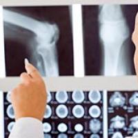 X-ray densitometry What is densitometry
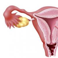 Is it possible to increase and increase the thickness of the endometrium to normal when planning a pregnancy for conception?
 Nutrition without diets from Cindy Crawford
 Hairpin interpretation of the dream book Why dream of buying a hairpin